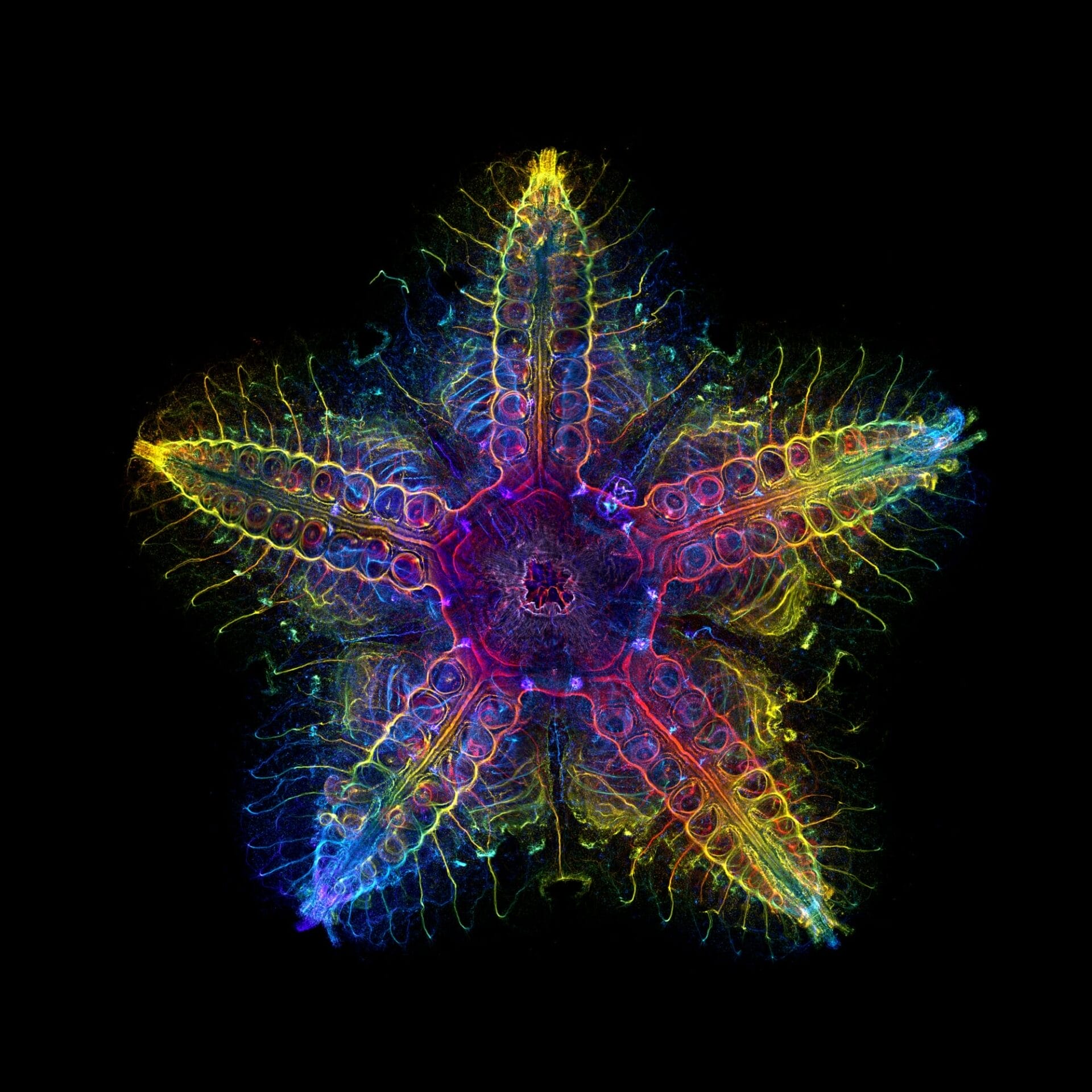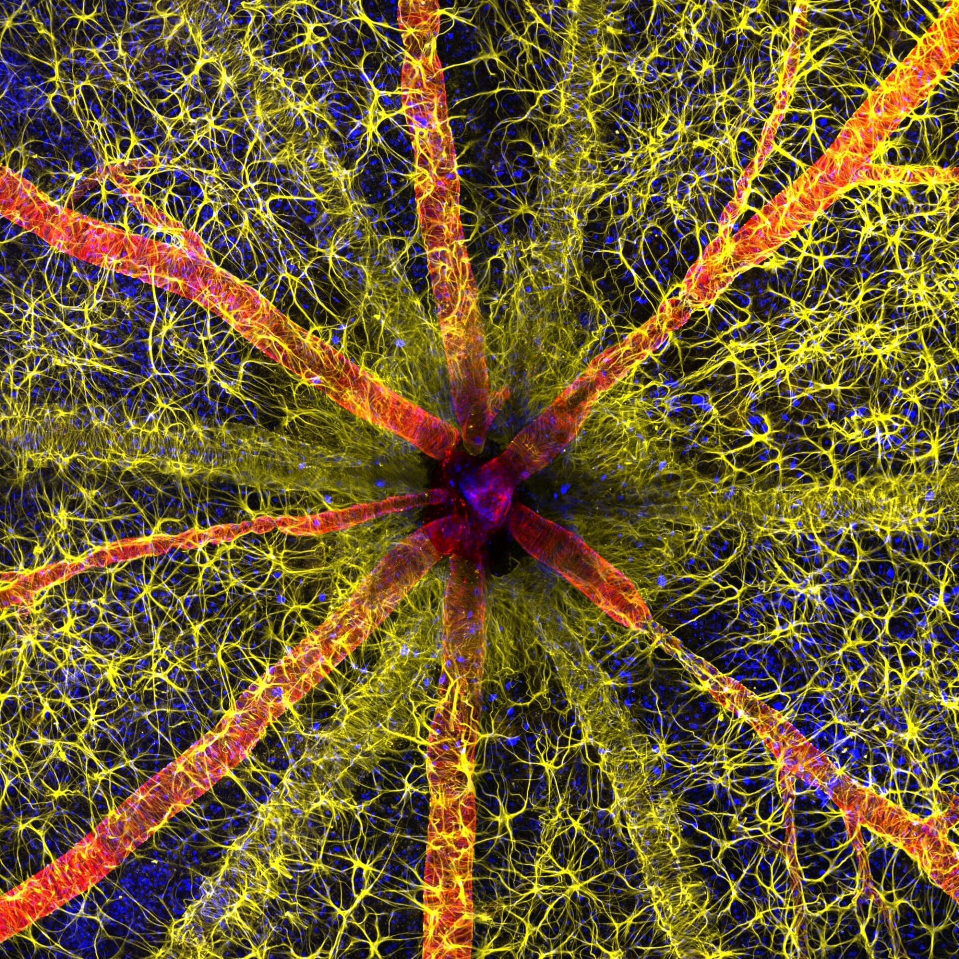
An Arresting Optic Nerve Tops the 2023 Nikon Small World Photomicrography Competition
One in five people with diabetes also suffers from retinopathy, a disease causing vision loss and blindness. Problems occur when high blood sugar causes cells to swell and leak, damaging the retina. Because symptoms aren’t always perceptible at early stages, though, many people aren’t diagnosed until the condition has already progressed.
Researchers like Hassanain Qambari and Jayden Dickson have been working toward visualizing the initial signs of retinopathy to aid in early detection, which they recently achieved in an electrifying image that won the 2023 Nikon Small World Photomicrography Competition. A starburst-like web of red and yellow fibers sprawl across the frame, which magnifies the intricacies of a rodent’s optic nerve and offers insight into its function. “The visual system is a complex and highly specialized organ, with even relatively minor perturbations to the retinal circulation able to cause devastating vision loss,” Qambari said. “I entered the competition as a way to showcase the complexity of retinal microcirculation.”
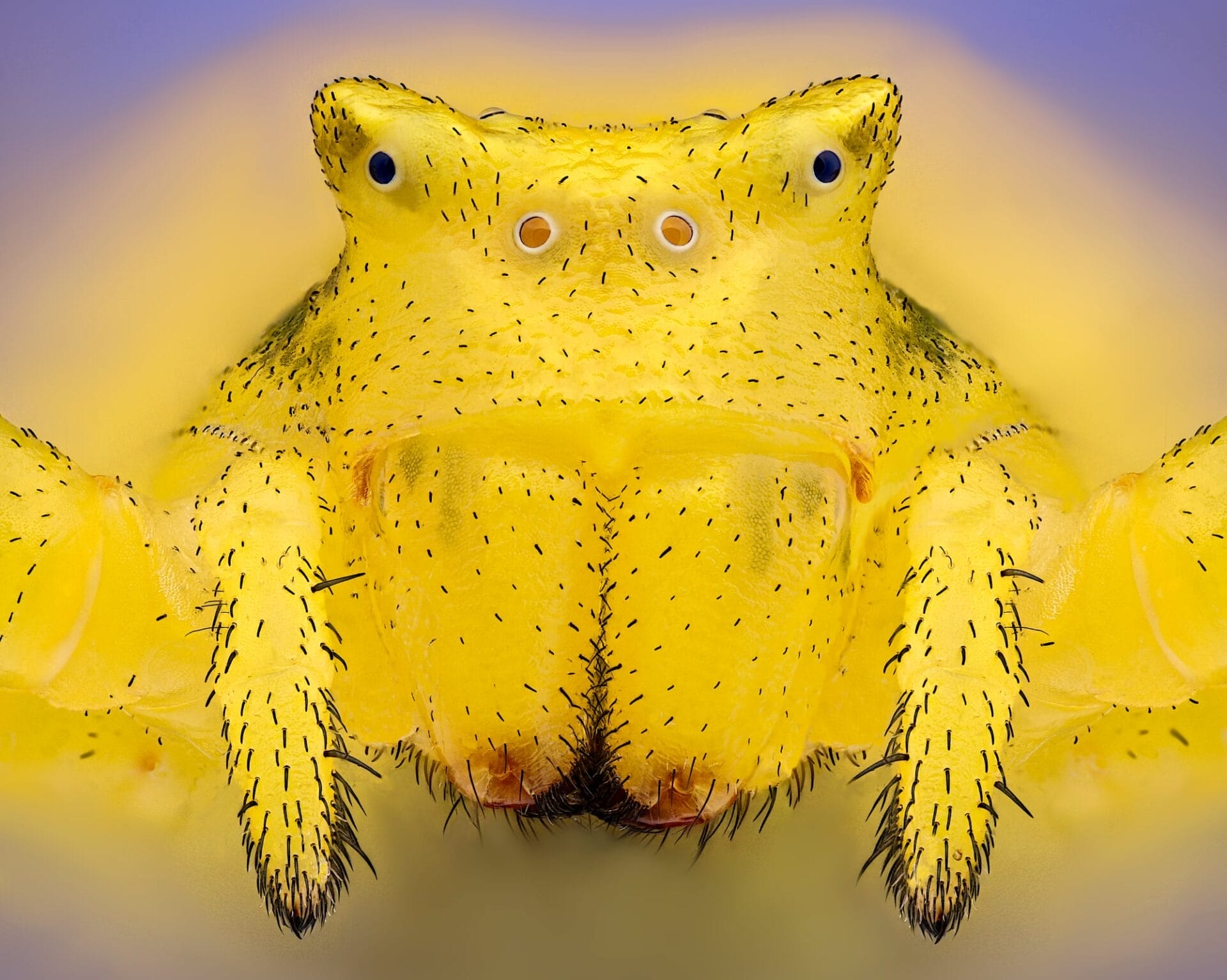
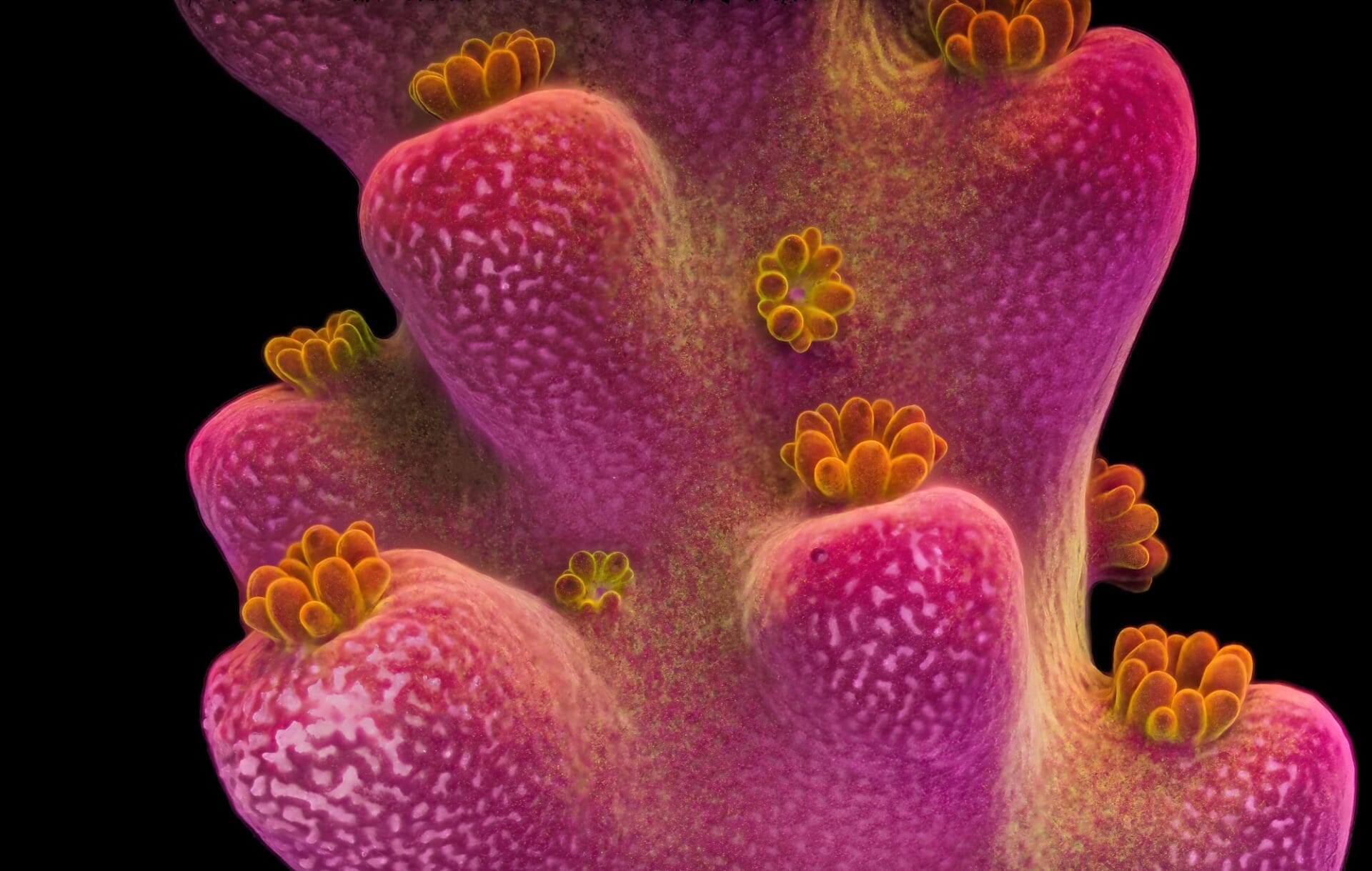
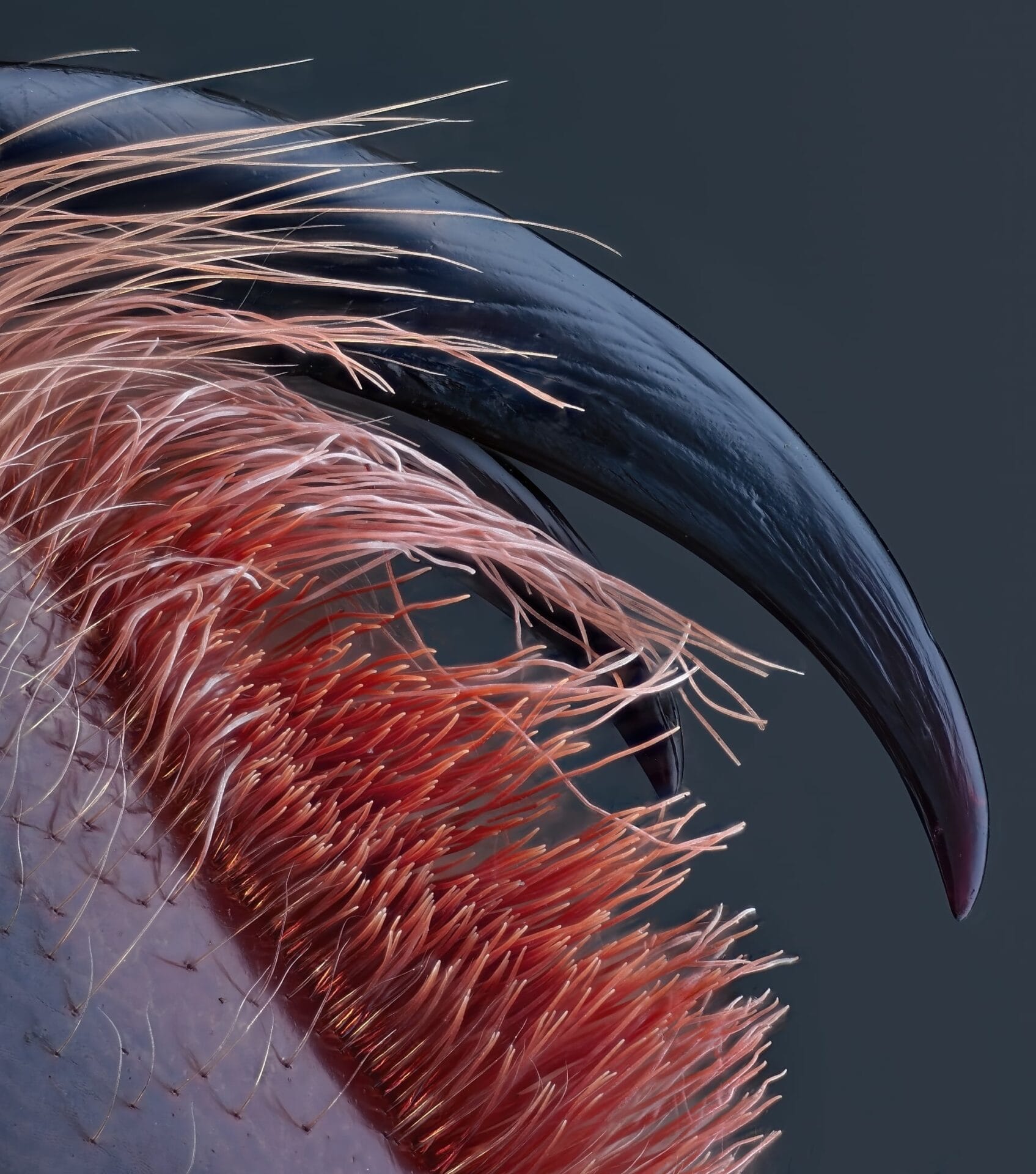
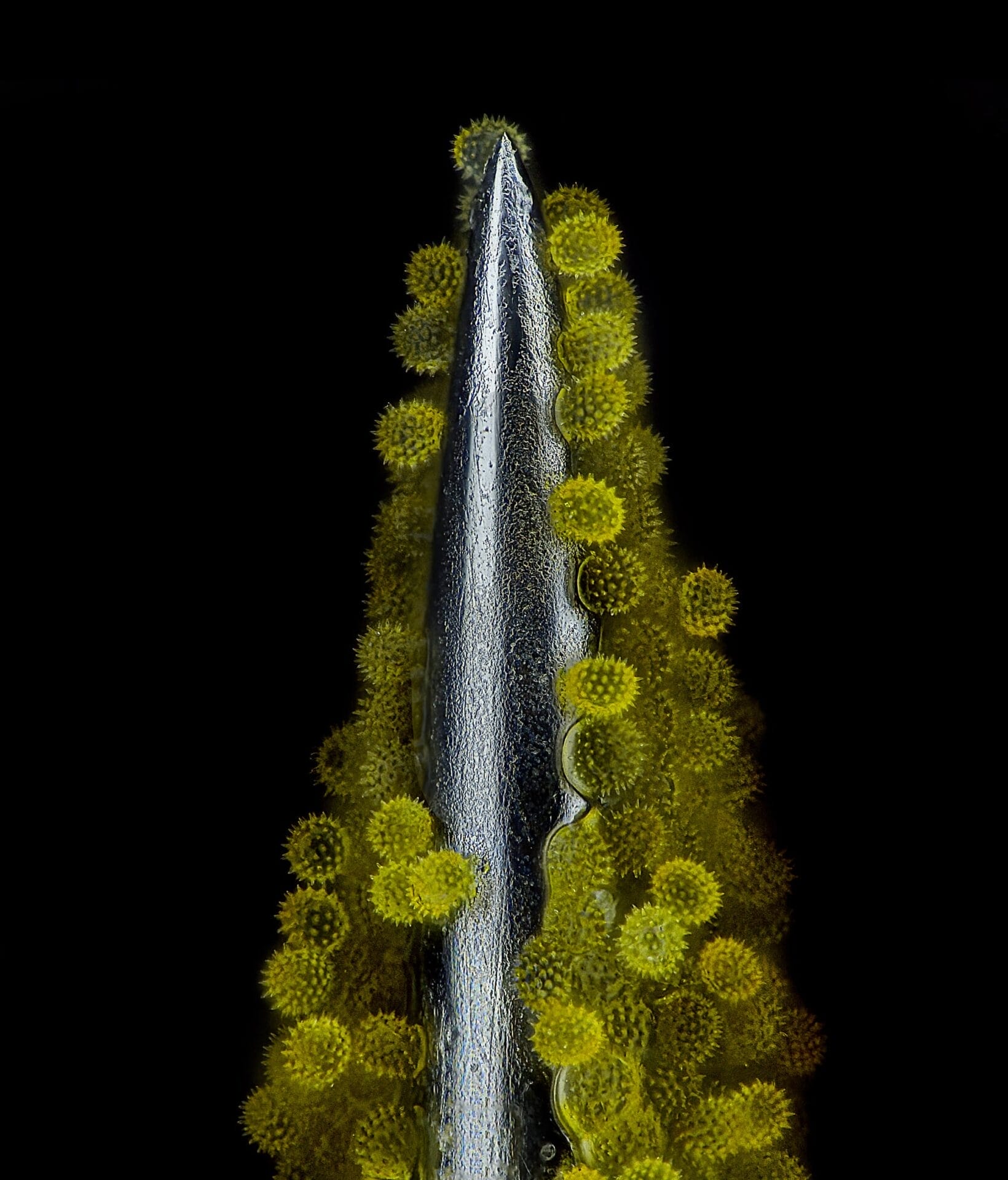
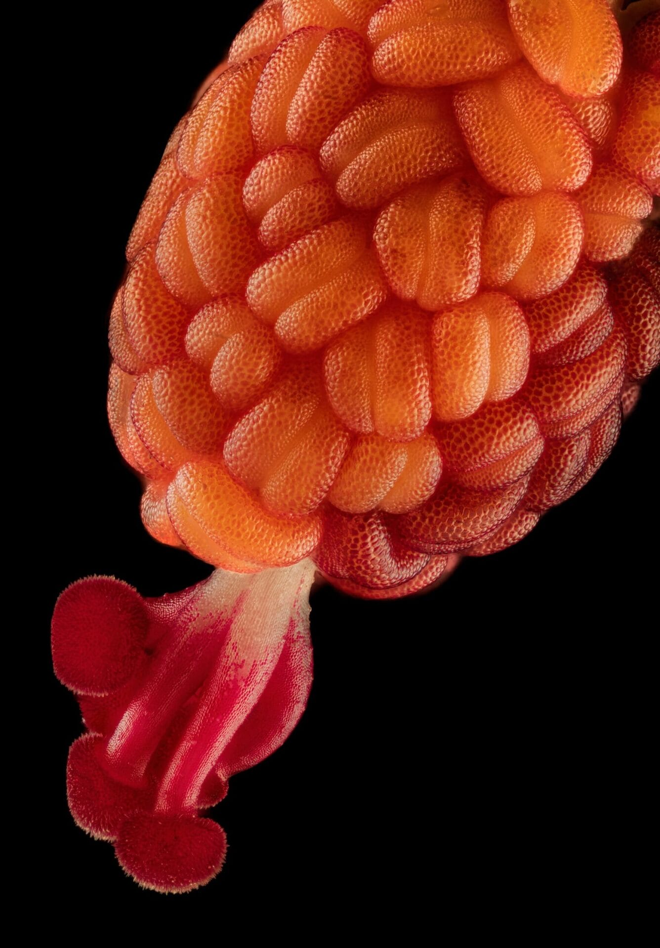
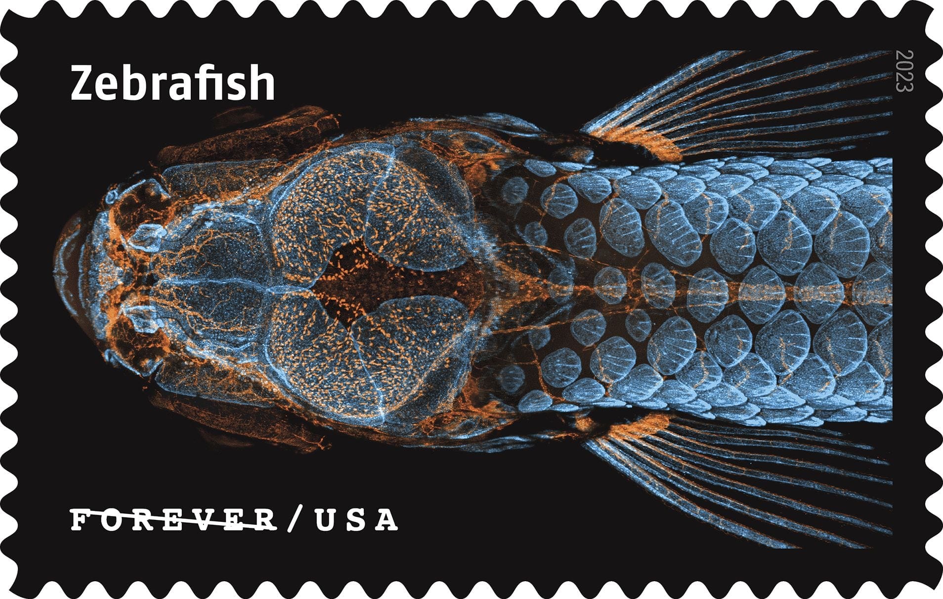
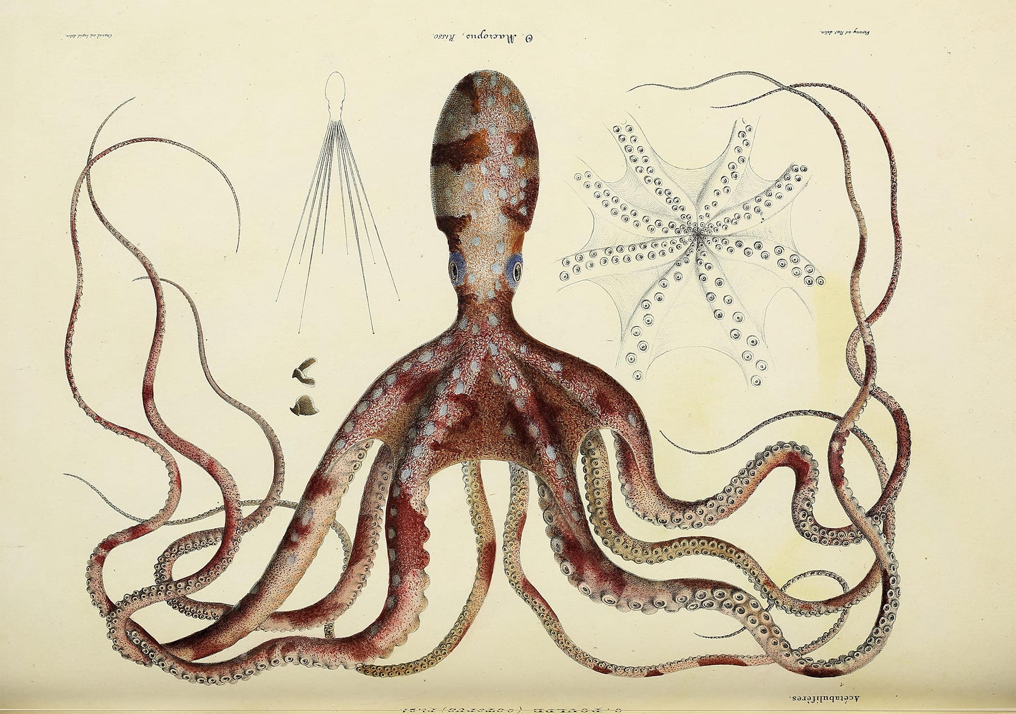
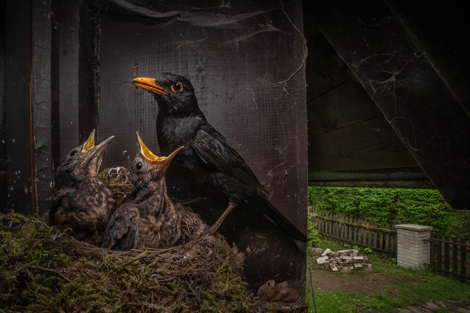
Other submissions to the 49th-annual contest include the venomous fangs of a tarantula, gelatinous slime molds, and spiky sunflower pollen stuck to an acupuncture needle. The 2023 competition garnered nearly 1,900 submissions from 72 countries, and you can see all of the winners on Nikon. It’s also worth taking a peek at the video segment of this year’s contest, which includes striking footage of neurons forming in an embryo.